Laparoscopic operation for retrocervical endometriosis
Deep infiltrative type of endometriosis remains urgent problem in gynecology. The notion “wide-spread endometriosis” implies massive and deep location of ectopic centre, usually involving Douglas’ cul-de-sac, tissues of anterior wall of rectum, posterior wall of vagina, uterus, sacrouteral ligaments. It promotes obliteration of space that is behind uterus and changing of its anatomy due to “soldering” of mentioned above formations. One or both walls of small pelvis are often involved into the pathological process in the area where ureters are and rectosigmoid part of the large intestine is located not far. Lesion of cystouterine sulcus, appendix, large intestine are met rarely.
Laparoscopic operation for external endometriosis demonstrates a surgeon’s experience.
Endometriosis centres affect different organs and tissues of the abdominal cavity and small pelvis. For the best results of treatment it is necessary to remove all the centres; there should be a radical approach to endometrioid lesion, but still preserving all organs and tissues, not doing harm to them, doctors should be especially careful with young patients who are going to become pregnant. That is why operation is efficient if it is performed on by an experienced surgeon not only in the field of gynecology, but in urology and proctology, as well. Unfortunately, if operation is performed on by a gynecological surgeon, according to legislation, he has no the right to perform on operations with involvement of large and small intestines, ureters, urinary bladder. That is why there is fear and unability to fulfil this complex task. It is good, if there is a surgeon in clinic who has got certificates to perform on operations in several fields of surgery, and if he can work in the field of laparoscopy. If not, centres are left, the operation is not radical. As I have mastered 4 specialities (surgery, gynecology, proctology, urology), it gives me a possibility to perform on such kinds of operations with a high quality and safely for patients within many years. Very often many patients come to me from the world around after 3-4 unsuccessful operations for deep infiltrative endometriosis with the lesion of many organs and systems. After successful treatment, even if it is stage 4 endometriosis, they enjoy a new quality of life and become happy mothers.
In order to determine the degree of lesion by endometriosis of the internal organs and to choose the correct tactics of surgical treatment, it is necessary to forward me (my e-mail is: puchkovkv@mail.ru puchkovkv@mail.ru  ) the full description of USI of organs of small pelvis, MRT data of small pelvis, the results of colonoscopy, to mention age and the main complaints. Then I will be able to give you an exact answer, related to your situation.
I have the experience of more than 4,000 successful laparoscopic operations for external endometriosis, including about 1,000 operations for wide-spread retrocervical endometriosis, stage 3-4.
The results of those laparoscopic operations are mentioned in monographs “Simultaneous Laparoscopic Operations in Surgery and Gynecology”, “Laparoscopic Operations in Gynecology” and in more than 60 scientific publications in different professional scientific publications in Russia and abroad. I have several patented techniques and diagnostic approaches, that are used in treatment of this category of patients. In 1997 for the first time in Russia I performed on simultaneous operation-laparoscopic extirpation of uterus and resection of large intestine for infiltrative endometriosis. Starting from that time the techniques and approaches to treatment of this category of patients have been changed greatly.
The main things that I use in treatment of infiltrative retrocervical endometriosis, I will write in sentences mentioned below.
- Retrocervical endometriosis is a surgical problem that can be solved only by properly performed on surgery. Hormones will not cure, but their proper use after operation can reduce the relapses of endometriosis within the range 7-3 %.
- The best access in treatment of retrocervical endometriosis is a laparoscopic one. Excellent visualization, precise technique of excision of centres within the boundaries of healthy tissues, even from the walls of cavities (large and small intestine, ureter and urinary bladder) without disturbing safety of lumen (the technique of shaving centres, improved by me) gives a possibility to achieve excellent functional results.
- Any disturbance of safety of serousmuscular covering of these centres should be accompanied by placing an atraumatic careful suture, using only synthetic absorbable thread.
- When you are revising organs of the abdominal cavity and small pelvis, you should additionally carefully examine diaphragm, uterine tubes, vermiform appendix and small intestine. Lesion of these organs is met very often in case of infiltrative endometriosis. Missing these centres, we leave endometriosis, that is able to cause complications-acute appendicitis, obstruction of small intestine, adhesion of organs of small pelvis, relapse of disease and infertility. If there is lesion of vermiform appendix, I perform on appendectomy, in case of lesion of small intestine, I use elaborated technique –“shaving” of the centres with wound stitching and preserving of intestine. The same thing is done, if there is localization of centres on uterine tubes. In case of diaphragm lesion I remove centres by low-temperature plasma, ‘Zering” device (Germany).
- Laparoscopic operation should be based on the exact command of anatomy of small pelvis, vascular and nervous structures. They all should be preserved. For improving of functional results of resection of large intestine I have put forward a new conception of performing on this operation laparoscopically. As a result, I remove only 3-4 cm of intestine ( not 15-25 cm as some colorectal surgeons do), preserving safe ampulla of rectum and normal function of defecation. This technique is described in detail below.
- For tissue dissection I use the contemporary electrosurgical complexes and platforms: ultrasonic scissors and an device of dosed electrothermal influence LigaSure” (Switzerland), that gives a possibility to perform on operation fast and without blood loss.
- In case of endometrioid cysts, especially if they are bilateral, we always investigate antimuller hormone (AMH) in the blood of a patient; reduction of it tells us that there is decrease of follicles in ovaries. In this situation, after cyst removal, I try not to coagulate the bed of ovary, but to use safe, but very efficient hemostatic “PerClot” (Italy); it is made of potato starch and is absorbed in 7 days. This approach helps to preserve follicles of ovaries, not reducing them by hemostasis of the bed of ovary by usual electrosurgical things-bipolar and monopolar coagulation.
- After operation we always use anticommissural barriers and gels for the sake of prophilaxis of adhesion development between fallopian tubes and adjacent organs and tissues.
Mentioned above surgical approaches give me a possibility to achieve excellent results in treatment of patients with infiltrative retrocervical endometriosis and infertility, to discharge them fast from hospital, to improve considerably their quality of life and restore their ability to become mothers.
Laparoscopic removal of retrocervical endometriosis with affected intestine. Surgical technique.
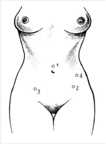
Figure 1. Points of introduction of trocars during hysterectomy, combined with anterior resection of the rectum (1–10 mm trocar; 2, 4–5 mm trocars; 3–12 mm trocar (K.V. Puchkov and co-authors, 2005)
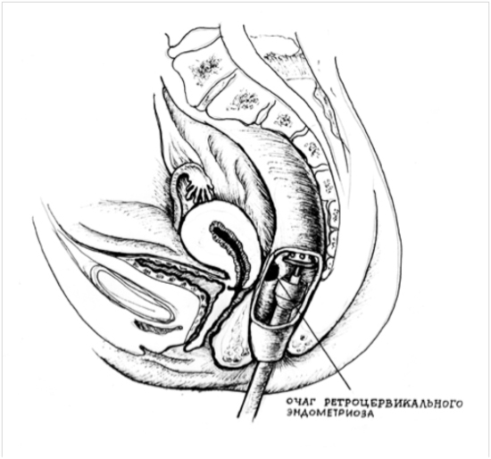
Figure 2. Transanal introduction of a circular stapler and its location over the endometrial focus (K.V. Puchkov and co-authors, 2005)
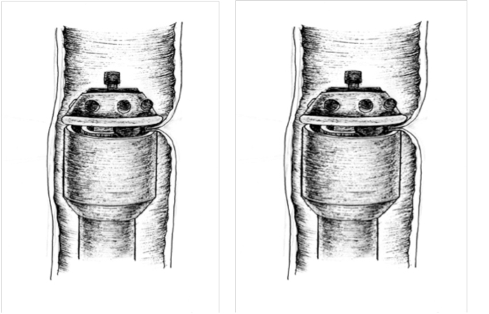
Figure 3. Segmental resection of the colon using the CEEA-33 circular stapler: 1) invagination of the endometriosis focus between the head and the anvil of the stapler; b) flashing stage (K.V. Puchkov and coauthors, 2005)
Operation depends on the degree of invasion and involvement into the pathological process adjacent organs; it is found out during preoperation rectoscopy and X-ray irigography, and intravenous pyelography or CT with the dye (contrast substance). A patient positioning is supinal with separated legs and a bent head (30 degrees).Through the umbilical access carboperitoneum is done, troacars and optics are introduced (fig.1).
Endometriosis of intestine, urinary bladder treatment, operation, laparoscopy points of introduction of troacars hysterectomy rectum
Under visual and tactile control abdomen is dissected on the border of unchanged tissue and firm endometrioid centre, that is exposed from lateral side in the area of sacrouterine ligaments, that very often presents considerable part of endometrioid infiltrate. Fibrously thickened part of sacrouterine ligament is dissected, including the area of its junction with the uterine isthmus. The use of scissors at this stage provides feeling of the consistency of dissecting tissues. “Crunching” means that this part is fibrously changed due to endometriosis; it testifies that affected tissues have been left there, i.e. that manipulations here have not been radical.
In case of extensive dissection of tissues in the area of lateral walls it is possible that retroabdominal fibrosis can develop; it should be born in mind in case of development of some deviations from the normal course of postoperation period. Preserving of vascular zones, located laterally to ureters, and the use of anticommissural barriers or gel in mentioned-above areas (Lyons T.L., 2001), can reduce the severity of this complication.
The volume of work on the urinary bladder depends on the depth of the lesion of the wall. Using a retractor, uterus is transferred into retroflexio position; and abdomen, covering urinary bladder, is opened. Excision of infiltrate by endoscissors within the boundaries of serous or serous-muscular layer requires placing single-layer suture, using absorbable thread. In case, if all the layers of walls of urinary bladder are involved into the process, the defect is closed by two- layer of interrupted suture, using “Vicryl” or “Monovicryl” 2-0 thread. Checking, whether suture is hemostatic or not, is done by introduction of methylene blue, using cystic catheter. Within 4-7 days after operation draining of the urinary bladder is done, using permanent catheter.
Excision of fibrous-changed centres of endometriosis, invading to the vaginal wall, is done by a monopolar electrode, using cutting current, when tissues are stretched by hard forceps. The defect of posterior wall of vagina is closed from the side of abdominal cavity by means of continuous suture, using “Vicryl” or “Monovicryl” 2-0 thread. Pneumoperitoneum is maintained by a special obturator or vaginal tampon. We often find hypertrophic tissues, not containing centres of endometriosis in the area of joining of the neck and vagina, sacrouteral ligaments and isthmus of uterus; they create difficulties while separating ectopic centres from this kind of tissues.
When performing on manipulations on the intestine that is involved into the endometrioid infiltrate, we always use intrauterine retractor and plastic intestinal retractor, giving a possibility to stretch tissues behind the uterine space and maximum visualize the area that is difficult to access. Fixation of uterus with the help of retractor in anteflexio position and the weight of rectum give us a possibility to get dosed stretching of tissues, necessary for safe exposure of endometrioid infiltrate.
Taking into consideration the possibility of wounding or arising of necessity to resect intestine, a patient who will be operated on for wide-spread endometriosis, requires a special preparation of intestine (fortrans or flit on the eve of operation).
The attempt to excise fibrous centre should be done only after the final exposure of intestine out of affected area until friable connective tissue of rectovaginal space appears. Tissues should be carefully separated bluntly, using aquadissector, or by a sharp method, using ultrasound scissors or an electrosurgical instrument.
For excision of endometrioid centre from the intestinal wall we use a monopolar needle electrode. In the mode of cutting serous-muscular layer on the border of unchanged and affected tissue is dissected. If only serous-muscular layer is involved into the pathological process, it is possible to excise the centre without opening the lumen of intestine (shaving). If before operation the symptom of invading of intestine is present, at first the endometrioid infiltrate should be separated from uterus.
In case of local lesion it is possible to do marginal resection of the large intestine. If there is intensive endometriosis of the large intestine with the deformity of walls, then a standard laparoscopic anterior resection of rectum is performed on or circular resection of colic intestine is done, and “end-to-end” anastomosis is formed by a circular stapler.

Patent. Method for temporary fixation of abdominal organs and small pelvis during laparoscopic operations
After marginal resection of intestinal wall the formed defect is stitched by manual atraumatic suture, using “Polysorb” thread 2-0, forming intracorporeal knots or by device method. The intestine should be compulsory stitched in transverse direction, it prevents development of stenosis and reduces the risk of failed suture. Defect stitching, using a linear stapler, is done in the following way: on the defect edges of intestine along transverse direction we apply 2-3 holders. We do traction, using holders, stitching by a linear stapler Endo TA-30, it makes anastomosis hermetic.
If the center is small, and it invades to mucous membrane, it is better to use the method of segmentary resection of the large intestine by a circular stapler, that combines simplicity and safety. After excision of the endometrioid centre from uterus 2 holders are applied onto the wall of affected intestine (proximally and distally from the centre) by atraumatic absorbable thread. A circulary stapler (the diameter is 33 mm) is introduced and fixed transanally so, that the centre is positioned between the head and anvil of the device (fig.2). A resecting part of intestine is invaginated to the axis of the device (fig.3a). For this the end of the thread (holder) is separated along the horizontal line perpendicular to the intestine, doing traction to the device axis. Stitching is done (fig. 3b), after that the dissected part of large intestine is removed alongside with the device. Stitching is physiological-there is no deformity of intestinal wall. And the diameter of dissected part can be 3.0 cm.
This method gives a possibility to decrease traumas, duration of operation, provides maximum grasp of changed intestinal wall. And in this case resection is done without opening the lumen of the large intestine, contamination by microflora of the abdominal cavity is absent, and it will promote to decrease the risk of arising of purulent-septic complications after operation.
Laparoscopic anterior resection of rectum is indicated, if endometriosis circularly grasps rectum (has the size not less than 3 cm), is located deep in small pelvis, often with some degree of obliteration or spread diffuse rectosigmoid lesion with invading to the wall, having clinical symptoms. If the surface of rigid intestine has a deepening in the centre, it is pathognomonic for extensive, deep endometrioid centre, that is not accessible for discus-shaped resection or excision up to the mucous membrane. Then we remove fat tissues (mesorectum) from the intestinal wall. At this area (the distal edge) it is transected by Endo GIA 30 (60) device at the distance of 1-2 cm from the affected edge.
Then the intestine is removed, using one of the described below methods.
The first variant of performing on operation for endometriosis. In the left iliac area (near troacar) the abdominal wall is dissected layer by layer. The loop of the sigmoid intestine is exteriorized into the wound at the area of the proximal edge of resection. The part of intestine that is necessary for anastomosis is exposed. At this area the intestine is transected between 2 forceps, and resected preparation is removed. Purse-string suture is placed onto the proximal edge. The head of a circular stapler is inserted into the lumen of intestine. Then purse-string suture is tightened. And the intestine is immersed into the abdominal cavity, and the anterior abdominal wall is stitched.
The second variant implies removal of intestine through the transverse incision above the pubic -3-4 cm.
The third variant is transvaginal removal of preparation. During this operation an affected segment of intestine is mobilized laparoscopically through the posterior colpotomy (that has been formed either during excision of the affected part of vagina, or has been done deliberately to remove it), the intestine with endometriosis is brought down up to the vestibule of vagina, where the part of intestine for anastomosis is exposed. At this area the intestine is transected between 2 forceps, and resected preparation is removed. Purse-string suture is placed onto the proximal edge. The head of a circular stapler is inserted into the lumen of intestine. Then purse-string suture is tightened. And the intestine is immersed into the abdominal cavity, and colpotomic opening is stitched by laparoscopic access.
After handling the stump of rectum by antiseptic solution the basic part of a circular stapler is introduced transanally; the sharp part of it perforates the stump in the mddle part of suture or in the area of angle, formed by Endo GIA-30 device. Then the head of an device is joined to the basic part of an device, stitching is done, and the instrument is removed.
Forming of coloanal or colorectal fistula in case of anterior resection of rectum with the help of a stitching device is considered to be the best method that reduces complication rate and preserving the function of anal sphincter.
It is worthy to mention that expanded resection of rectum, that surgeons usually perform on, is accompanied by development of different complications:
- injury of nerves ( anorgasmia, disuria);
- absence of rectal ampulla (disturbance of defecation, incontinence);
- worsening of life quality of a patient, non-satisfaction after operation.
These complications are related to the extensive dissection of tissues and removal of 12-25 cm of large intestine.
The decrease of mentioned-above complications are achieved in our clinic due to the following methods, elaborated by us:
- Exposure of intestine out of mesorectum, preserving visceral fascia (maximum preserving of all nerves).
- Performing on only resection of the changed part of intestine with maximum preserving of organ: the increase of the length of removed preparation is related to two factors: the increase of distance from the border of lesion and wrong choice of the way of removal of preparation from the abdominal cavity.
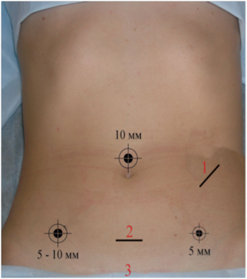
Figure 4. Accesses for removal of resected preparation of intestine depending on localization of endometrioid lesion (Puchkov K.V. et al, 2005).
During resection of intestine one should always try to preserve maximum each centimeter of an organ, it is achieved by dissecting of intestine in the distal direction not more than 1 cm from the pathological centre.
In order not to increase the length of resecting intestine, it is necessary to remove preparation from the abdominal cavity in a proper way, in the proximal direction, the line of dissection should also be not far than 1-2 cm from endometrioid infiltrate. It can be achieved by a special algorhythm of determining the point of removal of resected preparation of intestine, elaborated by us, it depends on localization of the endometrioid lesion (fig.4).
Access 1 – resection level is 18 cm and higher – removal of intestine through troacar access in the left iliac area (4-5 cm).
Access 2 - resection level – 12-20 cm – removal of intestine through incision according to Pfannenschtil (4-5 cm).
Access 3 - resection level is 8-12 cm – transvaginal or transanal removal of preparation.
If you are minding these simple conditions, it will give you possibility to remove not more than 4-7 cm of the large intestine. In this way you can avoid a lot of functional complications in young patients, having improved their quality of life.
The important condition of successful performing on of this stage of operation is to learn about arising injuries during operation. Cystoscopy and sigmoidoscopy after operation, as well as any other intraoperation investigations, necessary to testify presence or absence of disturbance of safety of intestine or urinary tract, are adequate control means for checking in case of intensive excision of tissue of these structures, that are not related to female reproductive system.
Thus, the surgical treatment of this category of patients is complex, long and requires a highly qualified surgeon, as its success is correlated to being radical in treatment.


