Cancer of uterus –surgical treatment, laparoscopic and laparotomic operations
Diagnostics
The diagnosis “cancer of uterus” is made up on the basis of a patient’s complaints and additional instrumental investigation - USI of organs of small pelvis and separate diagnostic abrasion under the guidance of hysterocervicoscopy (examination of uterine cavity, using special optic system). When hysterocervicoscopy is performed, localization of tumour is exactly determined, the degree of it extention and length, in case of histologic investigation of material, obtained in case of separate abrasion, and histological structure of tumour are determined; the degree of differentiation-it is very important to determine the risk of metastases and choosing the correct treatment plan. CT with dye (the contrast substance) and MRT give a possibility to determine the presence of metastases in lymphatic nodes and other organs.
Laparoscopy for cancer of uterus. The unique operation for uterine cancer, degree 1-2.
Indications for operation
Making up treatment plan depends on the degree of disease. If it is dergee1-2, the first and the main stage of operation is correct performing on operation in necessary oncological volume. In 90% of cases (if there are no contrindications in cardiovascular or pulmonary systems) operation can be performed laparoscopically, using several punches. Removal of uterus and appendages is done, and ileopelvic lymphadenectomy with removal of paravaginal fat is performed on. Laparoscopic access gives a possibility for a patient to get up and take food the next day after operation, and to be discharged from the clinic in 2-4 days. Absence of invasion and wound on the abdominal wall gives a possibility to start chemotherapy early in case of need.
In order to determine the degree of the disease in case of cancer of endometrium and to determine the degree of lesion of adjacent organs, and to choose the correct plan of the surgical treatment, it is necessary to forward me (my e-mail is: puchkovkv@mail.ru puchkovkv@mail.ru  ) a full description of USI of organs of small pelvis, MRT of small pelvis, performed with dye (a contrast substance), the results of hysteroscopy and histology, mention your age and complaints. Then I will be able to give you an exact answer about your situation.
If there are contrindications for intubational narcosis and pneumoperitoneum (that is compulsory in case of laparoscopy), then operation is performed on laparotomically, using peridural anesthesia.
After getting all data about disease stage (CT of lungs, CT or MRT of abdominal cavity and small pelvis, histology results after hysteroscopy, gynecological data about uterus and lymphatic nodes, IGI (immunohistological investigation) The further treatment plan is made up by an oncologist, a chemotherapeutist, a radiologist.
In our Swiss University clinic there is a possibility to hold consultation with all mentioned above specialists and after performing on operation and ask what to do next.
Peculiarity of my own tactics in surgical approach when treating cancer of uterine body
- Staging of oncological process and evaluation of somatic condition of a patient before operation give a possibility to choose properly the necessary volume and access of intereference (whether it will be laparoscopy or laparotomy):
- The use of laparoscopy gives a possibility to a patient to restore fast, without development of complications, and to start getting on time (in case of need) chemotherapy or radial therapy;
- Performing of the main stages of operation with the use of contemporary electrosurgical systems Force Triad “Liga Sure” (Switzerland) gives a possibility to ligate safely blood and lymphatic vessels, it reduces the operation time up to 1.5 hours and prevents lymph outflow after operation with lymphocele formation (accumulation of lymph in close space of the abdominal cavity);
- Removal of lymphatic nodes en block and removal of preparations in a special plastic container prevents contamination of the abdominal cavity by oncological cells;
- Restoration of the whole of ligamentary device of small pelvis at the end of operation by means of stitching of connective structures and vaginal walls layer by layer is done for the sake of prophilaxis of prolapse of genitals that may develop in future after operation- it considerably improves the life quality of patients;
- Adequate morphological mapping of lesion of uterus and lymphatic nodes in combination with instrumental pre-operation staging gives a possibility to choose the necessary scheme of further treatment of a patient.
Surgical treatment
The main technique of treatment of cancer of uterus is surgical one; it implies differentiated approach to use different kinds of operation. The operation volume can vary, depending on spreading process and risk of metastases development.
Laparoscopically I perform on the whole of the oncological volume, necessary for treatment of disease-starting from removal of uterus with its appendages to expanded extirpation of uterus with appendages, upper one-third of vagina, pelvic fat and lymphatic nodes.
My experience includes more than 300 laparoscopic operations for cancer of uterus with excellent and good results, they are mentioned in monographs and scientific publications. Annually I hold master-classes for gynecologists and oncologists about surgical treatment of cancer of uterus.
The technique of laparoscopic operation for cancer of uterus (endometrium).
I usually am to the right of a patient for fast and easier work, an assistant with a videocamera-by the head of a patient, the second assistant is between separated legs of a patient at the stage of panhysterectomy, he shifts uterus by means of a uterine manipulator, at the stage of lymphadenectomy he is to the left of a patient. The scheme of troacars introduction is shown at fig.1. 10-mm troacars are introduced above umbilicus and pubis, in iliac areas-5-mm troacars. One more 10-mm troacar is introduced into the left mesogastral area for introducing EndoBebcock forceps.
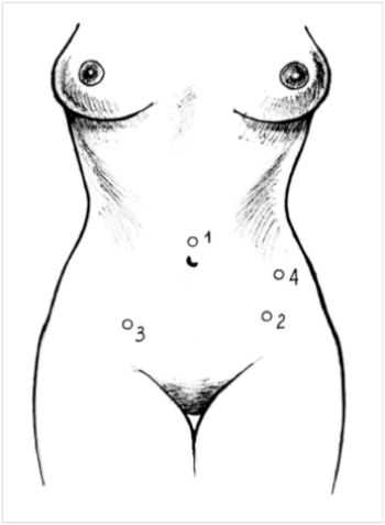
Figure 1. Points of introduction of trocars during uterus operations (K.V. Puchkov and co-authors, 2005)
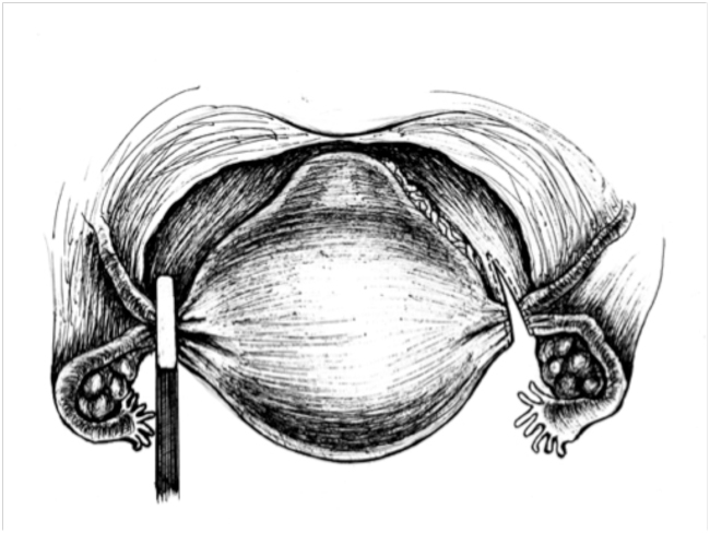
Figure 2. Mobilization of the ligament apparatus with the "LigaSure" apparatus (Switzerland) (K.V. Puchkov and co-authors, 2005).
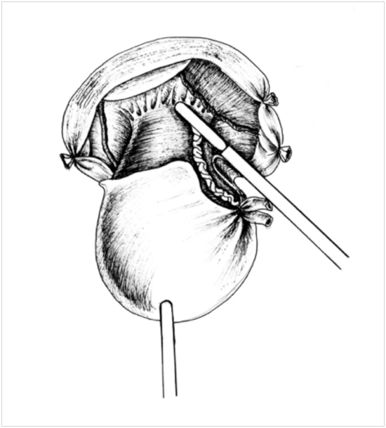
Figure 3. Coagulation and intersection of the uterine artery with the "LigaSure" apparatus (Switzerland) (K.V. Puchkov and co-authors, 2005).
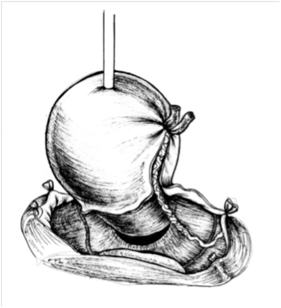
Figure 4. Opening the posterior vaginal fornix (K.V. Puchkov and co-authors, 2005).
Before introduction of uterine manipulator (with smooth cannula) I compulsory coagulate uterine tubes to prevent getting oncological cells from the uterine cavity into the abdominal cavity.
While performing laparoscopic examination, we learn the exact size of uterus, condition of appendages of uterus, adhesion process in the area of small pelvis, presence and location of cancinomatosis of abdomen, condition of liver and the round ligament.
At present in our clinic the technique of laparoscopic panhysterectomy (with broad dissection of the sacrouterine and cardinal ligaments) and lymphadenectomy is elaborated in detail in all stages:
- High coagulation and transection of the round uterine ligaments.
- Opening of the anterior leaf of the broad ligament.
- Exposure of the main anatomic orientation: the external and internal iliac arteries and veins, ureters, supracystic artery, obturator nerve.
- Coagulation and exposure of the infundibulopelvic ligament.
- Dissection of the posterior leaf of abdomen, putting ureter aside medially.
- Performing on lymphadenectomy with the coagulation of uterine vessels at some distance.
- Vesicovaginal dissection.
- Performing on anterior and posterior colpotomy.
- Removal of preparation of uterus and lymphatic nodes from the abdominal cavity in a plastic container (separately).
- Stitching and peritonization of the stump of vagina with the restoration of the pelvic floor layer by layer.
- Careful lavage and hemostasis of the operation area.
The technique of lymphadenectomy for cancer of uterus (endometrium)
I perform dissection of tissues up to the level of intraabdominal fascia-exact near bifurcation of aorta and up to the pre-renal leaf of retroabdominal fascia-laterally to intraabdominal fascia. I follow along the mentioned above fascia, I separate paraaortal, paracaval fat and fat in the area of bifurcation of aorta and iliac vessels from upwards downwards.
In this situation the lateral borders are ureters that are surrounded by fascial leaf (this fascial case is formed by two leaves of perinephric fascia ). Thus, the further dissection is performed along the intraabdominal fascia, moving retroabdominal fascia with ureters that are positioned there, laterally. The dorsal border of lymphodissection at this stage of operation is the lumbar muscle. Then lymphodissection in the area of aorta bifurcation and the inferior vena cava along iliac vessels is done.
The most difficult stage of pelvic lymphadissection is lateral lymphadenectomy-I have been teaching surgeons the basics of it for more than 20 years. At this stage one should be careful in order not to injure positioned more lateral relatively to external iliac artery genitofemoral nerve and iliac vein. The latter is usually in collapse condition due to the increased intraabdominal pressure because of pneumoperitoneum. I perform lymphodissection from obturator space in the following way. I expose ureter and lead it aside medially. Then I grasp by a hard forceps the edges of dissected abdomen along the external iliac artery more lateral relatively to ureter; by means of separation, using ultrasonic scissors, I remove fat in the direction to the external iliac vein.
An assistant leads aside external iliac vessels laterally and upwards with the help of an endoscopic retractor, in this way he opens the access to obturator areas. Obturator nerve with its vessels and the internal obturator muscle, that is an orientation of the depth of lymphodissection, is located backwards and medially. I remove fat, located under the external iliac vessels in the obturator area en block, preserving the obturator nerve; while doing it, I constantly perform traction of uterus upwards and to both sides.
Operation duration is about 40 minutes on both sides, if it is an experienced surgeon. The number of complications do not exceed 0.4%.
At all the key stages of oncological operations I use the contemporary techniques of miniinvasive surgery (ultrasonic scissors and an device of dosed electrothermal influence “LigaSure” (Switzerland); it gives a possibility to perform on operation fast and without blood loss. This energetic platform gives a possibility to make mobilization stage of uterus faster due to the automatic determining of tissue properties (tissue impedance); and depending on it, to provide dosed distribution of energy.You can read about this kind of energy in detail at our page ” The equipment for electrosurgery LigaSure” .The same thing regarding ultrasonic scissors; you can read about the advantages of it in the corresponding part of our site.
The use of these instruments gives a possibility to ligate safely both blood vessels and lymphatic ones, due to it operation time is reduced twice; and in postoperation period lymphorrhea and lymphocele are absent; but while using usual ways of lymphadenectomy and panhysterectomy, lymphocele and lymphorrhea are up to 20%.
For achieving maximum prophilaxis of postoperation prolapse we use the most contemporary synthetic stitching thread and restoration of ligamentary device of the pelvic area is done layer by layer.
For the sake of prophilaxis of adhesion disease we use the contemporary anticommissural barriers and gels, made in Switzerland and the U.S.A.
All operations, presented in this part, are considered to be complex and difficult in laparoscopic gynecology and oncology. The foundation of their successful performing on, definitely, is the proper work of the equipment and if members of the operation team get on with each other. Differentiated approach to select patients for laparoscopic panhysterectomy, proper use of different methods of hemostasis, dissection, joining tissues and surgical approaches give a possibility to increase safety and efficiency of operations.
In postoperation period all the patients are consulted by an oncologist and a chemotherapeutist to decide about the necessity of radial therapy or chemotherapy on the basis of exact morphological diagnosis and stage of the disease.
The accumulated great experience in treatment of patients for cancer of uterine body gives a possibility to mention about the following advantages of the laparoscopic access:
Minimum traumas of the anterior abdominal wall, it promotes to activate patients early, many of them have the risk factor of thromboembolism and postoperation pneumonia, postoperation rehabilitation is much more easier, a restoration period is shorter;
Better visualization of the surgical site (as compared to laparotomy) that gives a possibility to perform operations carefully and radically, as well as to find out metastases into the lymphatic nodes and to remove them during operation;
On average duration of operation is 1-1.5 hours. Next day patients are able to get up and take thin food. In 2-3 days they are discharged from the clinic. In 7 days they have control examination, using a gynecologist’s chair. In case of need USI of small pelvis organs is done. The data of histological investigation are compulsory discussed with other doctors (an oncologist, a chemotherapeutist, a radiologist) to decide the further treatment.


