Esophageal Achalasia
The Notion of Esophageal Achalasia, the Spreading of the Disease
Spreading of this disease in Europe is 1 person for 100,000 of population, in the structure of all diseases of esophagus its spreading is within the range 2-25%.
Esophageal achalasia is non-ability of the exit part of esophagus to relax as a result of congenital absence of specific nervous fibres in the intermuscular layer. In case of esophageal achalasia esophagus becomes deprived of innervation at some small area. On the background of it its increased sensibility to gastrin and cholinomimetics (hormones of local regulation) is formed. Due to it, within some period of time, the area of stable stenosis of esophagus in front of pylorus of stomach is formed. In case of this disease food gets into stomach not in a reflectory way (due to swallowing), but due to mechanical opening of cardia on the background of the hydrostatic pressure, formed by the column of thin food, accumulated in the esophagus by that time. At first the muscular force of the superior parts of esophagus can manage pushing food through that narrow area, then the muscle becomes weaker, and esophagus is expanded and becomes S-shaped.

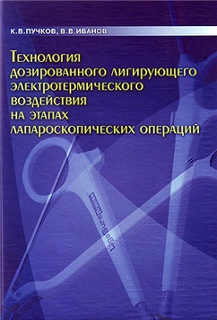
K.V. Puchkov, V.V. Ivanov and co-authors. Technology of metered ligation electrothermal effects at the stages of laparoscopic operations: Textbook, 2005

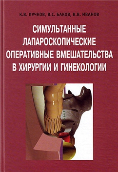
K.V. Puchkov, V.S., Bakov, V.V., Ivanov. Simultaneous laparoscopic surgeries in surgery and gynecology: Textbook, 2005

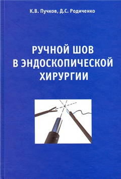
K.V. Puchkov, D.S. Rodichenko. Manual suture in endoscopic surgery: Textbook, 2004
Classification of Esophageal Achalasia
In the development of the disease they distinguish four stages:
- The first (functional) stage – temporary disorder of passing food via the cardial sphincter due to stopping of relax during food swallowing;
- The second stage is accompanied by moderate dilatation of the esophageal wall;
- The third stage is characterized by a stable dilatation of esophagus and stable stenosis in its inferior part due to scar changes
- The fourth stage is characterized by marked clinical presentation that is developed on the background of S-shaped dilatation of esophagus and complications, such as esophagitis, periesophagitis, fibrous mediastenitis, chronic bronchitis, chronic pneumonia, and atelectasis in lungs (on the background of constant night reflux of contents of esophagus and aspiration of food into respiratory tract).
Symptoms and Clinical Presentation of Esophageal Achalasia
The first complaints of patients are as follows: disturbance of swallowing (dysphagia), lump in the throat, then pain behind the sternum develops, coughing, food regurgitation.
Development of esophageal achalasia is rather slow, but the degree of the main symptoms of the disease is constantly increasing. Dysphagia occurs while taking different food, both thin food and thick one, as a rule, in several seconds after starting to swallow. And food is not in the throat, but in the thorasic cavity, that is why feelings are as if of retrosternum localization.
Regurgitation is reflux of food from inferior into superior parts (into the oral cavity). Reflux is increased, when a person bends after taking food, and at night, when a patient is in a horizontal position. Food remnants often get into the respiratory tract, and complications and clinical manifestations develop.
Odynophagia is a pain in the thorasic cavity, it is related to the spasm of smooth musculature of esophagus and overfilling of its lumen by food.
When the fourth degree starts to develop, stagnation symptoms occur due to long time keeping swallowed food in the esophagus ( offensive breath is in the oral cavity, sensation of nausea, foul-smelling eructation). On the background of it much lactic acid is formed; that is why heartburning and inflammation of the esophageal walls develop- esophagitis – it can be of erosive and ulcerative kind.
Diagnostics of Esophageal Achalasia
Diagnostics of this disease is based on the data, obtained while asking a patient questions ( a patient’s complaints), physical examination of a patient and the results of the instrumental investigation.
The main X-ray symptom of the esophageal achalasia is a stenosis of the distal part of the esophagus that has even clearly-marked contours, and a dilated area of GIT is located above it. Above the constricted part there is often hanging of esophageal walls-the symptom of ”feather for writing”.
Fibroendoscopic method gives a possibility to evaluate visually the condition of the therminal part of the esophagus and gastroesophageal isthmus. In most cases it is possible to pass the fibroscope via the constricted part of esophagus, it proves the fact that there are functional changes, and most probably, it is an esophageal achalasia.
Manometry of esophagus permits to evaluate objectively movable activity of the organ and the work of the inferior and superior sphincters of esophagus. Disturbance of constriction abilitiy of esophagus and cardial sphincter develop earlier as compared to the development of the clinical symptoms of the disease. That is why manometry is the main method of early diagnostics of the disease, at this time you cannot find symptoms of the disease, using X-ray or FGS.
In order to find out esophageal achalasia and to choose the correct method of the surgical treatment, it is necessary to forward me the full description of gastroscopy, X-ray of esophagus and stomach, using barium, USI of organs of the abdominal cavity, mention your age and the main complaints. My e-mail is as follows:
puchkovkv@mail.ru
puchkovkv@mail.ru  In case, when there are contradictory facts (X-ray, FGS), it is necessary to perform manometry of the esophagus. Then I will be able to give the exact answer relatively to your situation.
Treatment of Esophageal Achalasia. Medicament Therapy and Cardiodilatation
It is necessary to mention that pharmtherapy (calcium channel-blocking agents, nitrates, myotropic spasmolytics, painkillers) are efficient only during the first stage, when mild and rare dysphagia is present.
The acknowledged method of treatment is an endoscopic gullet bougienage, aimed to dilate the constricted area, but, unfortunately, rupture of tissues takes place in this case, and rough scars are formed in this zone, and it makes the situation worsened in future. Efficiency after bougienage is not long, repeated manipulations are required, and after 4-5 procedures a patient does not feel better. In this situation a patient is told to be operated on for this problem-esophagomyotomy (it is an incision of a part of esophagus up to the submucous layer without opening the lumen of an organ), it gives a possibility to restore a normal swallowing, and a patient will have a normal mode of life.
The endoscopic method of dilatation can be useful for aged patients and for those ones who have had prolapse of the disease after operation. Here the use of the endoscopic method implies those patients for whom operation is contraindicated or it is difficult to perform on this operation for some category of patients.
One of the miniinvasive methods of treatment of esophageal achalasia is an endoscopic intrasphincter injection of botulotoxin A.It is considered to be an alternative efficient version for those patients who have absolute contraindications to endoscopic hydro- or pneumatic cardiodilatation and surgical interference, esp. for aged patients and for those ones, who have severe accompanying pathology of cardiovascular and bronchopulmonary systems. Treatment by botulotoxin A is safe, but one should bear in mind that the effect in the inferior esophageal sphincter is short-2-3 months, that is why it should be done several times.
It is very important! The peculiarity of approach to choose the method of treatment of patients with the esophageal achalasia is an early determination of indications to laparoscopic operations, while there is no complication development and useless waste of time for unnecessary conservative treatment.
Early operations give good results, as in the constricted parts there are no scars due to the previous cardiodilatation, and the muscular force of the esophageal wall is not exhausted yet. But even in case of dilated esophagus good results are possible in case if a doctor uses an adequate laparoscopic technique.
Surgical Methods of Treatment of Esophageal Achalasia.
A lot of surgical methods have suggested to treat esophageal achalasia, but only those ones are used that deal with extramucous cardiomyotomy. One of the main techniques is an extramucous cardioplasty according to E.Heller. The essence of it is a longitudinal incision of the muscular layer of the distal part of esophagus along its anterior wall-the length is 8-10 cm. And myotomy should embrace not only the stenosis area and the cardial part of stomach, but partially dilated part of esophagus.
According to data, mentioned in scientific literature, good results after this kind of surgical interference have been registered in 90% of cases. The shortcoming of this technique is related to the fact that the mucous membrane of esophagus at the long distance (8-10 cm x 1 cm) remains unprotected. That is why at the areas of incision of the muscular layer of esophagus diverticulum can be formed, as well as scars, deforming cardia, promoting relapse of the disease.
The rate of lethal outcome after operation according to E.Heller is on average about 1.5% (but some authors say, it is 4%). The main cause of death is lesion of the mucous membrane of esophagus-missed cases-it promotes development of mediastinitis (inflammation of mediastinum fat), pleuritis (inflammation of pleura and peritonitis (inflammation of abdomen). Trauma of the mucous membrane is found out in 6-10 % of all the operations, that is why for the sake of prophilaxis of severe complications it is important to revise this area carefully. If a surgeon finds out the injury of the mucous membrane, it should be compulsory stitched. Unsatisfactory results are also related to disturbance of the cardial sphincter and development of peptic reflux-esophagitis. And due to the possibility of joining and fusion of the incised walls of the muscular layer relapse of the disease develops, dysphagia and disturbance of emptifying of esophagus take place.
It is important! For reducing lethal outcome and the number of postoperation complications it is necessary to cover surgical defect of the esophageal wall by own tissues of a patient, but, unfortunately, it is rarely done by surgeons as this stage is not easy to perform.
Nowadays there are different ways to cover the mucous membrane. For this purpose omentum can be used, the anterior wall of stomach, part of esophagus. It is important to know that versions of closure of defect of the muscular layer of esophagus, using some synthetic material, is not desirable because pressure sore can develop at this area.
Laparoscopy in Surgical Treatment of Esophageal Achalasia and its Complications
Laparoscopic operations for diseases of esophagus and stomach are distinguished by their delicacy and being functional.The increased image on the screen permits to visualize properly all details of anatomical formations in this area for careful operating- vagus nerve, gastric vessels and fascial space. Laparoscopic access gives a possibility to reduce operation time, shortens postoperation rehabilitation period and makes it easier, and promotes reducing the number of complications and gives a good cosmetic effect.
We have tried to make the technique of laparoscopic operation for esophageal achalasia perfect, having reduced the number of relapses up to 0.2%, and the number of complications-up to 1%.
My approach to surgical treatment of patients with esophageal achalasia is the following:
- Early determination for indications to operation; there is no need to wait for development of complications and to waste time for useless conservative treatment;
- To use only laparoscopic access: better visualization, less traumas, better cosmetic effect, fast recovery.
- Performing on an extramucous cardioplasty with the compulsory excision of muscular and scar layers at the distance of 8-10 cm; it provides the guaranteed liquidation of stenosis;
- For the sake of prophilaxis to prevent development of complications because of the open mucous layer, it is necessary carefully to revise that area; in case of need stitching should be done and compulsory covering of the anterior wall of stomach;
- For the sake of prophilaxis of disease relapse (fusion of the incised muscular layer) covering of the area of the anterior wall of stomach should be done, with the compulsory stitching of it along the perimeter of the defect of the mucous membrane, using the technique, elaborated by Professor K.V.Puchkov; placing atraumatic suture, using synthetic resorbable thread;
- For the sake of prophilaxis of insufficient cardial sphincter (because of the incision of the muscular layer) and development of peptic esophagitis strenhthening of this zone by the anterior wall of stomach (fundoplication-120 degrees) should be performed on;
- When exposing esophagus and stomach, I use the modern device of dosed electrothermal ligating of tissues “LigaSure” (U.S.A.), it gives a possibility to ”weld” vessels without injuring surrounding structures and, using a thin electrode, to dissect the muscular layer of esophagus without injury of the mucous membrane;
- The technique, elaborated by me, gives a possibility not to leave a transgastral probe (it makes the postoperation period easier); a patient can take thin food next day after operation.
As a result of using our method the overwhelming majority of our patients (97%) tell about good results. And the positive result is testified by the results of fibroesophagogastroduodenoscopy, X-ray investigations and esophagomanometry. According to the results of manometry we have achieved reducing of basal tonus of esophagus in all the operated patients. Besides, values of esophageal contractility are considerably improved, as well as some increase of cardia length due to forming of fundoplication cuff takes place. According to values, obtained within a 24-hour intraesophageal pH-metry, pathological gastroesophageal reflux ( regurgitation of gastric contents into esophagus) after operation according to E.Heller in combination with esophagofundoplication according to Dore in our modification, is registered at the physiological level.
My experience includes more than 600 operations for hiatal hernia and reflux-esophagitis and more than 100 operations for esophageal achalasia. They are mentioned in 3 monographs and in more than 60 scientific publications in different professional editions in Russia and abroad.
After operation 3-4 incisions remain on the skin of abdomen, having the length 5-10 mm. Patients start getting up and taking water at first day; next day after operation they are allowed to take warm thin food. They leave the clinic in 1-3 days depending on the severity of the disease. A patient can resume his job in 2-3 weeks. He should keep a strict diet within 1.5-2 months, and a mild diet-within 6 months. Then a patient will be able to proceed to his usual mode of life-without tablets and keeping diet.
If a patient wishes, he can have total investigation before operation to choose the method of operation and to determine the optimum tactics of treatment.


